

[Nov-2024] SPI Dumps are Available for Instant Access from Exam-Killer
Study resources for the Valid SPI Braindumps!
ARDMS SPI Exam Syllabus Topics:
| Topic | Details |
|---|---|
| Topic 1 |
|
| Topic 2 |
|
| Topic 3 |
|
| Topic 4 |
|
| Topic 5 |
|
NEW QUESTION # 58
Which factor affects temporal resolution?
- A. Time gain compensation
- B. Overall gain
- C. Log compression
- D. Display depth
Answer: D
Explanation:
Temporal resolution refers to the ability of an ultrasound system to distinguish between events occurring closely in time. It is primarily affected by the frame rate, which is the number of frames displayed per second. One of the main factors that influence the frame rate is the display depth. The deeper the imaging depth, the longer it takes for the ultrasound pulses to travel to the target and back, thus reducing the frame rate and temporal resolution. Shallower imaging depths allow for higher frame rates and better temporal resolution.
Reference:
ARDMS Sonography Principles and Instrumentation (SPI) Exam Study Guide
"Diagnostic Ultrasound: Principles and Instruments" by Frederick W. Kremkau
NEW QUESTION # 59
What is an advantage of power Doppler over color Doppler?
- A. Diminished flash artifact
- B. Increased frame rate
- C. Accurate velocity information
- D. Less angle dependent
Answer: D
Explanation:
Power Doppler, unlike color Doppler, is less angle dependent because it detects the strength of the Doppler signal rather than the velocity of the blood flow. This means it is more sensitive to detecting low-velocity flow and flow in smaller vessels, regardless of the angle between the ultrasound beam and the flow direction. Color Doppler provides information on flow direction and velocity but is highly dependent on the angle of insonation, making it less reliable when the angle is suboptimal.
Reference:
ARDMS Sonography Principles and Instrumentation guidelines
Zwiebel, W. J., & Pellerito, J. S. (2017). Introduction to Vascular Ultrasonography. Elsevier.
NEW QUESTION # 60
What is the primary factor that improves lateral resolution?
- A. Frame rate
- B. Propagation speed
- C. Frequency
- D. Beamwidth
Answer: D
Explanation:
Lateral resolution refers to the ability of the ultrasound system to distinguish two structures that are side by side, perpendicular to the direction of the sound beam. This resolution is primarily improved by reducing the beamwidth. A narrower beamwidth allows for better differentiation between adjacent structures, enhancing the lateral resolution. Higher frequency transducers can also help achieve a narrower beamwidth, but beamwidth is the primary factor.
Reference:
ARDMS Sonography Principles & Instrumentation Guidelines
Hagen-Ansert SL. Textbook of Diagnostic Ultrasonography. 8th ed. St. Louis, MO: Mosby; 2017.
NEW QUESTION # 61
Which describes the reflected frequency when a reflector is moving toward the sound source?
- A. Unchanged
- B. Decreased
- C. Increased
- D. Attenuated
Answer: C
Explanation:
When a reflector (such as red blood cells) is moving toward the sound source, the frequency of the reflected sound waves increases. This phenomenon is known as the Doppler effect. The frequency shift occurs because the motion of the reflector compresses the sound waves, leading to a higher frequency than the emitted frequency. This increased frequency is what the Doppler ultrasound system detects and uses to calculate the velocity of the moving reflector.
Reference:
ARDMS Sonography Principles and Instrumentation guidelines
Hoskins, P. R., Thrush, A., Martin, K., & Whittingham, T. A. (2010). Diagnostic Ultrasound: Physics and Equipment.
NEW QUESTION # 62
According to Poiseuille's law, a change in which parameter would have the greatest influence on blood flow?
- A. Vessel radius
- B. Length of vessel
- C. Viscosity of the fluid
- D. Pressure gradient
Answer: A
Explanation:
According to Poiseuille's law, the flow rate of a fluid through a vessel is directly proportional to the fourth power of the vessel's radius. Therefore, a small change in the radius of the vessel has a much larger effect on blood flow compared to changes in pressure gradient, length of the vessel, or viscosity of the fluid.
Reference:
ARDMS Sonography Principles and Instrumentation guidelines
Poiseuille's law in medical physics and hemodynamics literature.
NEW QUESTION # 63
Which statement describes the purpose of using a spectral Doppler wall filter?
- A. To eliminate the lower velocity signals
- B. To clean up the audio signals
- C. To eliminate the higher velocity signals
- D. To widen the area in which the Doppler shift is sampled
Answer: A
Explanation:
The purpose of using a spectral Doppler wall filter is to eliminate lower velocity signals. Wall filters are designed to remove low-frequency Doppler shifts caused by the motion of the vessel walls or surrounding tissues, which are generally of no diagnostic value. By eliminating these lower velocity signals, the wall filter helps to clean up the Doppler signal and reduce clutter, allowing for a clearer and more accurate display of blood flow velocities.
Reference:
ARDMS Sonography Principles and Instrumentation (SPI) Exam Study Guide
"Diagnostic Ultrasound: Principles and Instruments" by Frederick W. Kremkau
NEW QUESTION # 64
Which feature is a characteristic of continuous wave Doppler?
- A. Aliasing
- B. Range specificity
- C. Dedicated transmit and receive crystals
- D. Low thermal index
Answer: C
Explanation:
Continuous wave Doppler uses two crystals - one for transmitting and one for receiving ultrasound waves continuously. This allows for the measurement of high velocities without aliasing, a common limitation in pulsed wave Doppler. However, continuous wave Doppler does not have range specificity, meaning it cannot precisely determine the depth from which the Doppler signal is returning.
Reference: ARDMS Sonography Principles and Instrumentation, Chapter on Doppler Ultrasound.
NEW QUESTION # 65
What happens to the amount of attenuation if the path length is doubled?
- A. Doubled
- B. Quartered
- C. Quadrupled
- D. Halved
Answer: A
Explanation:
Attenuation in ultrasound is directly proportional to the path length. If the path length is doubled, the amount of attenuation is also doubled. Attenuation refers to the reduction in the amplitude and intensity of the ultrasound wave as it travels through tissue, primarily due to absorption, reflection, and scattering. The relationship is linear, so doubling the distance the sound wave travels will result in twice the amount of attenuation.
Reference:
ARDMS Sonography Principles and Instrumentation guidelines
Kremkau, F. W. (2015). Diagnostic Ultrasound: Principles and Instruments. Elsevier.
NEW QUESTION # 66
What is the primary reason to use compression?
- A. Adjust the contrast resolution
- B. Improve the axial resolution
- C. Increase line density
- D. Reduce the focal region
Answer: A
Explanation:
Compression in ultrasound imaging adjusts the range of grayscale displayed, affecting the contrast resolution.
This function allows sonographers to enhance the differentiation between structures of varying echogenicities.
By modifying the contrast resolution, sonographers can better visualize subtle differences in tissue composition and improve the diagnostic quality of the images.
Increasing contrast resolution is particularly important in differentiating between fluid-filled cysts and solid masses. Reference:
ARDMS Sonography Principles and Instrumentation guidelines on image processing and contrast resolution.
NEW QUESTION # 67
How is intensity of an ultrasound beam measured?
- A. Hydrophone
- B. Autocorrelation
- C. Reynold's number
- D. Doppler equation
Answer: A
Explanation:
The intensity of an ultrasound beam is measured using a hydrophone. A hydrophone is a specialized device that detects and measures the acoustic pressure of the ultrasound waves in water or tissue-mimicking materials. It is highly sensitive and can measure the variations in pressure, which are used to calculate the intensity and other acoustic parameters of the ultrasound beam.
Reference:
ARDMS Sonography Principles and Instrumentation guidelines
Hoskins, P. R., Thrush, A., Martin, K., & Whittingham, T. A. (2010). Diagnostic Ultrasound: Physics and Equipment.
NEW QUESTION # 68
Which setting can be increased to correct for clutter artifact when using pulsed-wave Doppler?
- A. Pulse repetition frequency (PRF)
- B. Sample volume
- C. Wall filter
- D. Doppler gain
Answer: C
Explanation:
The wall filter, also known as the high-pass filter, is used in Doppler ultrasound to remove low-frequency signals, which are typically associated with clutter artifacts. Clutter artifacts can be caused by tissue motion or vessel wall movement, and they appear as low-frequency signals that can obscure the desired blood flow signals. By increasing the wall filter setting, these low-frequency signals are filtered out, thus reducing the clutter artifact and providing a clearer depiction of the blood flow.
Reference:
American Registry for Diagnostic Medical Sonography (ARDMS). Sonography Principles and Instrumentation (SPI) Examination Review Guide.
NEW QUESTION # 69
Which resolution capability is most affected by spatial pulse length?
- A. Temporal
- B. Axial
- C. Lateral
- D. Elevational
Answer: B
Explanation:
Axial resolution refers to the ability to distinguish two structures that are close to each other along the path of the ultrasound beam.
Spatial pulse length (SPL) is the distance over which one pulse occurs, and it directly affects axial resolution.
Shorter SPL improves axial resolution because it allows better differentiation of closely spaced structures.
The axial resolution is improved by increasing the frequency of the transducer, which shortens the wavelength and hence the SPL. Reference:
ARDMS Sonography Principles and Instrumentation guidelines on resolution parameters and their impact on image quality.
NEW QUESTION # 70
Which ultrasound adjustment allows for an increased frame rate in color flow Doppler?
- A. Using multiple focal zones
- B. Decreasing depth
- C. Using continuous wave Doppler
- D. Increasing sector scan width
Answer: B
Explanation:
Frame rate in color flow Doppler is influenced by several factors, including the imaging depth. Decreasing the depth reduces the time it takes for sound waves to travel to the imaging area and back to the transducer. This allows for more frames to be captured per second, thereby increasing the frame rate. Higher frame rates improve temporal resolution, making it easier to visualize moving structures.
Reference:
ARDMS Sonography Principles & Instrumentation Guidelines
Hagen-Ansert SL. Textbook of Diagnostic Ultrasonography. 8th ed. St. Louis, MO: Mosby; 2017.
NEW QUESTION # 71
What occurs when the pulse repetition frequency is less than twice the Doppler shift frequency?
- A. Spectral broadening
- B. Range ambiguity
- C. Aliasing
- D. Propagation speed artifact
Answer: C
Explanation:
Aliasing occurs in Doppler ultrasound when the pulse repetition frequency (PRF) is less than twice the Doppler shift frequency (Nyquist limit). When this condition is met, the Doppler signals are not sampled frequently enough to accurately measure the frequency shifts, resulting in the misrepresentation of the flow velocities. This causes the aliasing artifact, where high-velocity flows are displayed incorrectly as wrapping around the baseline, leading to potential diagnostic errors.
Reference: ARDMS Sonography Principles and Instrumentation (SPI) Review, Doppler Artifacts section.
NEW QUESTION # 72
Which adjustment can maintain the same frame rate when the depth is increased?
- A. Decrease image width
- B. Decrease persistence
- C. Increase frequency
- D. Increase number of focal zones
Answer: A
Explanation:
When the depth of imaging is increased, the time it takes for the ultrasound pulses to travel to and from the deeper structures also increases, which can reduce the frame rate. To maintain the same frame rate, one effective adjustment is to decrease the image width. Narrowing the image width reduces the number of scan lines required to create each frame, allowing the system to maintain a higher frame rate despite the increased depth.
Reference:
ARDMS Sonography Principles and Instrumentation guidelines
Kremkau, F. W. (2015). Diagnostic Ultrasound: Principles and Instruments.
NEW QUESTION # 73
Which artifact causes a reflector to be improperly positioned on the display?
- A. Enhancement
- B. Range ambiguity
- C. Acoustic shadowing
- D. Speckle
Answer: B
Explanation:
Acoustic Shadowing: This artifact occurs when a structure absorbs or reflects most of the ultrasound waves, causing a shadow behind the structure. It does not cause improper positioning of a reflector on the display.
Speckle: This is a form of noise in ultrasound imaging that appears as granular texture. It can affect image quality but does not lead to improper positioning of reflectors.
Enhancement: This artifact occurs when the area behind a weakly attenuating structure appears brighter. It affects the brightness of the image but does not affect the position of reflectors.
Range Ambiguity: This occurs when an echo is received after the next pulse has been sent out, causing the reflector to be placed at an incorrect depth on the display. This is because the system assumes the echo came from the most recent pulse.
Reference:
"Ultrasound Physics and Instrumentation" by Frank Miele
ARDMS Sonography Principles and Instrumentation study materials
NEW QUESTION # 74
Which artifact may be caused by incorrect color Dopplergain setting?
- A. Aliasing
- B. Bleed/Blossoming
- C. Twinkle
- D. Clutter/Haze
Answer: B
Explanation:
Incorrect color Doppler gain settings can cause the artifact known as bleed or blossoming. When the color Doppler gain is set too high, it can cause the color signal to "bleed" outside the actual boundaries of the blood vessel, leading to an overestimation of the area of flow. This artifact makes it appear as though the blood flow extends beyond the true vessel walls, which can obscure the accurate interpretation of the Doppler image.
Reference:
ARDMS Sonography Principles and Instrumentation (SPI) Exam Study Guide
"Diagnostic Ultrasound: Principles and Instruments" by Frederick W. Kremkau
NEW QUESTION # 75
What is a potential negative consequence of using a high wall filter?
- A. Too much noise may appear on the image
- B. Aliasing could occur
- C. Penetration is reduced
- D. Desired signal may be eliminated
Answer: D
Explanation:
A high wall filter is used in Doppler ultrasound to eliminate low-frequency signals that may be attributed to vessel wall motion or other low-velocity flows. However, if the wall filter is set too high, it can inadvertently eliminate desired low-frequency Doppler signals that represent real blood flow, particularly in smaller vessels or those with slower flow velocities. This results in a loss of valuable diagnostic information.
Reference: ARDMS Sonography Principles and Instrumentation (SPI) Review, Doppler Ultrasound section.
NEW QUESTION # 76
Which action may reduce the number of lines in a frame without a loss of temporal resolution?
- A. Reducing the frame rate
- B. Narrowing the field of view
- C. Decreasing the display depth
- D. Decreasing the transducer frequency
Answer: B
Explanation:
Narrowing the field of view reduces the number of scan lines that need to be processed per frame. This allows the ultrasound system to maintain or even increase the frame rate without compromising temporal resolution. Temporal resolution, which refers to the system's ability to depict motion accurately, is directly related to the frame rate. Reducing the field of view ensures fewer lines are needed to create each image, thus preserving the frame rate and temporal resolution.
Reference:
ARDMS Sonography Principles and Instrumentation guidelines
Kremkau, F. W. (2015). Diagnostic Ultrasound: Principles and Instruments.
NEW QUESTION # 77
Which artifact displays reflectors more shallow than their actual position?
- A. Section thickness
- B. Mirror image
- C. Range ambiguity
- D. Ring-down
Answer: C
Explanation:
Range ambiguity artifact occurs when echoes from one pulse are received after the next pulse has been emitted, leading to the incorrect placement of echoes at shallower depths than their true location. This artifact typically happens when the PRF is set too high, causing the ultrasound system to interpret delayed echoes as coming from the current pulse rather than the previous one. This results in reflectors appearing closer to the transducer than they actually are.
Reference:
ARDMS Sonography Principles & Instrumentation Guidelines
Kremkau FW. Sonography Principles and Instruments. 9th ed. Philadelphia, PA: Elsevier; 2016.
NEW QUESTION # 78
What is the relationship between overall gain and image brightness?
- A. There is no relationship between overall gain and image brightness
- B. The lower the overall gain, the brighter the image
- C. The higher the overall gain, the brighter the image
- D. The higher the overall gain, the darker the image
Answer: C
Explanation:
Overall gain in ultrasound refers to the amplification of all the received echo signals. Increasing the overall gain amplifies the signals, making the entire image brighter. Conversely, decreasing the overall gain reduces the signal amplification, resulting in a darker image. Overall gain adjustment affects the entire image uniformly, unlike time gain compensation (TGC), which adjusts the gain at different depths independently.
Reference:
ARDMS Sonography Principles and Instrumentation (SPI) Exam Study Guide
"Diagnostic Ultrasound: Principles and Instruments" by Frederick W. Kremkau
NEW QUESTION # 79
Which factor does a string phantom evaluate?
- A. Intensity values
- B. Flow velocity
- C. Slice thickness
- D. Two-dimensional resolution
Answer: B
Explanation:
A string phantom is designed to evaluate the accuracy of Doppler ultrasound systems, specifically in measuring flow velocity. It consists of a moving string or filament that mimics blood flow within a vessel. By using this phantom, sonographers can assess how accurately the ultrasound system can detect and measure the speed of the moving target. This helps in calibrating and verifying the performance of Doppler systems, ensuring they provide accurate flow velocity readings in clinical practice.
Reference:
American Registry for Diagnostic Medical Sonography (ARDMS) Sonography Principles and Instrumentation study materials.
Textbook of Diagnostic Sonography by Hagen-Ansert, S. L. (latest edition).
NEW QUESTION # 80
What adjustment is needed to optimize the color in the image below?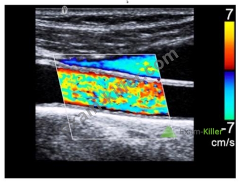
- A. Increase wall filter
- B. Increase pulse repetition frequency
- C. Decrease persistence
- D. Decrease gain
Answer: B
Explanation:
Increasing the pulse repetition frequency (PRF) helps to optimize the color Doppler imaging by reducing aliasing.
Aliasing occurs when the PRF is too low to accurately sample the rapid blood flow velocities, leading to incorrect color representation.
By increasing the PRF, the system can more accurately measure higher velocities without distortion, improving the overall quality of the color Doppler image. Reference:
ARDMS Sonography Principles and Instrumentation guidelines on Doppler imaging and techniques to reduce aliasing.
NEW QUESTION # 81
......
Updated SPI Tests Engine pdf - All Free Dumps Guaranteed: https://www.exam-killer.com/SPI-valid-questions.html
Latest ARDMS SPI SPI Actual Free Exam Questions: https://drive.google.com/open?id=1qfn24RzxBht98lPzeozfi-Z66n7oFHIY
