![]()
Get Ready with SPI Exam Dumps (2026)
Realistic SPI Dumps are Available for Instant Access
ARDMS SPI Exam Syllabus Topics:
| Topic | Details |
|---|---|
| Topic 1 |
|
| Topic 2 |
|
| Topic 3 |
|
| Topic 4 |
|
| Topic 5 |
|
NEW QUESTION # 19
What produces increased attenuation within soft tissue?
- A. Higher intensity of the ultrasound beam
- B. Lower frequency of the ultrasound beam
- C. Higher frequency of the ultrasound beam
- D. Lower intensity of the ultrasound beam
Answer: C
Explanation:
Attenuation refers to the reduction in the intensity of the ultrasound beam as it travels through tissue. Higher frequency ultrasound beams experience more attenuation because they are absorbed and scattered more than lower frequency beams. This is due to the fact that higher frequency waves have shorter wavelengths and interact more with the small particles in tissues, causing greater energy loss.
References:ARDMS Sonography Principles and Instrumentation, Chapter on Ultrasound Physics and Instrumentation.
NEW QUESTION # 20
What information does the ultrasound system calculate to display color flow?
- A. Minimum velocity of flow
- B. Peak velocity of flow
- C. Peak Doppler frequency
- D. Mean Doppler frequency
Answer: D
Explanation:
Color flow Doppler imaging displays the mean Doppler frequency shift, which represents the average velocity of blood flow within a sample volume. The ultrasound system uses autocorrelation to process Doppler signals and compute the mean frequency shift. This provides a color-coded map of blood flow velocities, allowing for visualization of flow direction and speed. The mean Doppler frequency is displayed as different colors, with each color representing a range of velocities.
References:
ARDMS Sonography Principles & Instrumentation Guidelines
Kremkau FW. Sonography Principles and Instruments. 9th ed. Philadelphia, PA: Elsevier; 2016.
NEW QUESTION # 21
What is an advantage of using pulsed wave Doppler as compared to using continuous wave Doppler?
- A. Higher echo sensitivity
- B. Ability to select sample depth
- C. Decreased display of aliasing
- D. Improved temporal resolution
Answer: B
Explanation:
Comprehensive and Detailed Explanation From Exact Extract:
The key advantage of pulsed wave Doppler is range resolution, meaning the operator can select a specific depth (sample volume) for measuring velocities. Continuous wave Doppler does not provide this capability, as it samples velocities along the entire beam path.
According to sonography instrumentation reference:
"Pulsed wave Doppler allows selection of sample volume depth, providing range resolution which continuous wave Doppler lacks." Therefore, the correct answer is B: Ability to select sample depth.
NEW QUESTION # 22
How is intensity of an ultrasound beam measured?
- A. Doppler equation
- B. Hydrophone
- C. Autocorrelation
- D. Reynold's number
Answer: B
Explanation:
The intensity of an ultrasound beam is measured using a hydrophone. A hydrophone is a specialized device that detects and measures the acoustic pressure of the ultrasound waves in water or tissue-mimicking materials. It is highly sensitive and can measure the variations in pressure, which are used to calculate the intensity and other acoustic parameters of the ultrasound beam.
Reference:
ARDMS Sonography Principles and Instrumentation guidelines
Hoskins, P. R., Thrush, A., Martin, K., & Whittingham, T. A. (2010). Diagnostic Ultrasound: Physics and Equipment.
NEW QUESTION # 23
Which index is related to the likelihood of cavitation?
- A. Mechanical
- B. Temporal
- C. Acoustical output
- D. Thermal
Answer: A
Explanation:
The Mechanical Index (MI) is related to the likelihood of cavitation, which is the formation of gas bubbles in a liquid due to the low-pressure regions of the ultrasound wave. MI is a parameter that predicts the potential for mechanical bioeffects, including cavitation. A higher MI indicates a greater likelihood of cavitation occurring. It is calculated based on the peak negative pressure and the frequency of the ultrasound wave.
Reference:
ARDMS Sonography Principles and Instrumentation guidelines
Kremkau, F. W. (2015). Diagnostic Ultrasound: Principles and Instruments.
NEW QUESTION # 24
Which artifact is caused by defects in the crystals of the transducer?
- A. Mirror image
- B. Dropout
- C. Side lobe
- D. Ringdown
Answer: B
Explanation:
Comprehensive and Detailed Explanation From Exact Extract:
Defects in transducer crystals result in missing or weakened signals along the beam path produced by those elements, creating dropout. In array transducers, dropout typically appears as vertical or horizontal dark zones depending on which elements are affected.
According to sonography instrumentation reference:
"Crystal failure results in areas of signal dropout directly beneath the defective elements due to loss of transmitted or received signals." Therefore, the correct answer is D: Dropout.
-
NEW QUESTION # 25
Which is a method to reduce noise?
- A. Increase frequency
- B. Decrease depth
- C. Increase persistence
- D. Decrease beam width
Answer: C
Explanation:
Persistence is a form of temporal averaging where consecutive frames are averaged to reduce random noise, resulting in a smoother image. Increasing persistence effectively reduces noise by averaging out transient noise artifacts while preserving the true signal. This improves image quality, although it may also reduce the temporal resolution, making it less suitable for rapidly moving structures.
Reference:
ARDMS Sonography Principles & Instrumentation Guidelines
Hedrick WR, Hykes DL, Starchman DE. Ultrasound Physics and Instrumentation. 4th ed. Philadelphia, PA: Elsevier Saunders; 2005.
NEW QUESTION # 26
How can the spectral Doppler mirroring seen in this image be eliminated?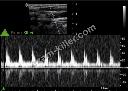
- A. Decrease wall filter.
- B. Increase pulse repetition frequency (PRF).
- C. Decrease Doppler gain.
- D. Increase dynamic range.
Answer: C
Explanation:
Spectral Doppler mirroring, also known as crosstalk, occurs when the Doppler signal appears on both sides of the baseline. This can be caused by excessively high Doppler gain, which amplifies the signal and creates artificial mirror images. Decreasing the Doppler gain reduces the signal amplitude, thereby minimizing the mirroring artifact.
Reference:
ARDMS Sonography Principles and Instrumentation guidelines
Hoskins, P. R., Thrush, A., Martin, K., & Whittingham, T. A. (2010). Diagnostic Ultrasound: Physics and Equipment.
NEW QUESTION # 27
How can spectral Doppler cross talk be reduced?
- A. By decreasing the Doppler gain
- B. By adjusting the Doppler baseline
- C. By increasing the Doppler pulse repetition frequency (PRF)
- D. By decreasing the Doppler wall filter
Answer: A
Explanation:
Comprehensive and Detailed Explanation From Exact Extract:
Cross talk (spectral mirror artifact) appears as a duplication of the spectral waveform above and below the baseline. One primary cause is excessive Doppler gain, which amplifies weak signals and noise symmetrically. Reducing gain minimizes this artifact.
Principles and Instrumentation state:
"Cross talk in spectral Doppler may result from high gain settings. Lowering the gain can reduce or eliminate this artifact." PRF adjustments affect aliasing, not cross talk.
Baseline shifts affect waveform positioning.
Wall filter affects low-frequency signals but not cross talk.
Therefore, the correct answer is A: By decreasing the Doppler gain.
NEW QUESTION # 28
Which color Doppler control allows for reassignment of red and blue to represent flow direction?
- A. Color priority
- B. Color gain
- C. Color invert
- D. Color threshold
Answer: C
Explanation:
Comprehensive and Detailed Explanation From Exact Extract:
In color Doppler imaging, the default color map assigns red and blue to represent flow direction relative to the transducer (usually red toward, blue away). The color invert control reverses this assignment.
According to sonography Principles and Instrumentation:
"The color invert control reverses the baseline of the color map, swapping red and blue assignments for flow direction."
* Color priority adjusts the overlay of color vs grayscale.
* Color gain controls amplification of the Doppler signal.
* Color threshold sets minimum amplitude to display color.
Therefore, the correct answer is D: Color invert.
-
NEW QUESTION # 29
What method can be used to resolve aliasing artifact?
- A. Decreasing the pulse repetition frequency
- B. Adjusting the output power
- C. Using a higher frequency transducer
- D. Using continuous wave Doppler ultrasound
Answer: D
Explanation:
Comprehensive and Detailed Explanation From Exact Extract:
Continuous wave (CW) Doppler can measure very high velocities without aliasing because it does not have a Nyquist limit like pulsed-wave Doppler.
Principles and Instrumentation state:
"Aliasing is eliminated in continuous wave Doppler since it does not rely on sampling and has no upper velocity limit." Output power (A) affects signal strength, not aliasing.
Higher frequency (B) increases aliasing susceptibility.
Decreasing PRF (C) actually worsens aliasing.
Therefore, the correct answer is D: Using continuous wave Doppler ultrasound.
-
NEW QUESTION # 30
Which parameters determine the propagation speed of sound in a medium?
- A. Frequency and impedance
- B. Elasticity and density
- C. Amplitude and impedance
- D. Intensity and density
Answer: B
Explanation:
The propagation speed of sound in a medium is determined by the medium's elasticity and density. Elasticity refers to the ability of the medium to return to its original shape after deformation, while density is the mass per unit volume of the medium. The speed of sound increases with higher elasticity and decreases with higher density. This relationship is described by the equation#=##v=#E, where#vis the propagation speed,#Eis the elasticity (or modulus of elasticity), and##is the density.
References
* ARDMS Sonography Principles and Instrumentation (SPI) Exam Study Guide
* "Diagnostic Ultrasound: Principles and Instruments" by Frederick W. Kremkau
NEW QUESTION # 31
Which artifact causes a simple cyst to appear to contain debris?
- A. Slice thickness
- B. Enhancement
- C. Refraction
- D. Range ambiguity
Answer: A
Explanation:
Comprehensive and Detailed Explanation From Exact Extract:
Slice thickness (elevational resolution artifact) occurs when the ultrasound beam partially includes surrounding tissue above or below the scan plane, falsely appearing as internal echoes within an otherwise anechoic cyst.
Principles and Instrumentation:
"Slice thickness artifact occurs when off-axis echoes are included in the imaging slice, falsely creating internal echoes in cystic structures." Refraction (A) causes displacement.
Enhancement (B) causes posterior brightening.
Range ambiguity (D) produces incorrect depth placement.
Therefore, the correct answer is C: Slice thickness.
-
NEW QUESTION # 32
Which control should a sonographer use to change contrast resolution?
- A. Output power
- B. Dynamic range
- C. Reject
- D. Gain
Answer: B
Explanation:
* Reject: This control eliminates low-level noise and weak signals, affecting image quality but not primarily used for contrast resolution.
* Output Power: This adjusts the intensity of the transmitted ultrasound waves but does not directly change contrast resolution.
* Gain: This control amplifies all signals equally, affecting brightness but not specifically the contrast resolution.
* Dynamic Range: Adjusting the dynamic range changes the range of grayscale that the ultrasound system displays, which directly affects the contrast resolution by altering how many shades of gray are visible between the black and white extremes.
References:
"Understanding Ultrasound Physics" by Sidney K. Edelman
ARDMS Sonography Principles and Instrumentation study materials
NEW QUESTION # 33
Which technique averages individual frames together to improve the image?
- A. Harmonic imaging
- B. Coded excitation
- C. Compression
- D. Persistence
Answer: D
Explanation:
Persistence is a technique used in ultrasound imaging that averages individual frames together to improve the overall image quality. This process helps to reduce noise and improve the signal-to-noise ratio, leading to clearer and more stable images. By averaging multiple frames, transient artifacts are minimized, and the continuity of structures is better visualized. Persistence is particularly useful in imaging static or slow-moving structures.References:
* ARDMS Sonography Principles and Instrumentation guidelines
* "Ultrasound Physics and Technology: How, Why and When" by M. Evans, C. Archer, and K. Weston
NEW QUESTION # 34
Which situation occurs when the incident angle of a sound beam is adjusted to be perpendicular to a soft tissue interface?
- A. Cavitation
- B. Reflection
- C. Refraction
- D. Range ambiguity
Answer: B
Explanation:
Comprehensive and Detailed Explanation From Exact Extract:
Reflection is maximized when the ultrasound beam strikes a tissue interface at 90 degrees (perpendicular). This angle provides optimal return of echoes for imaging.
According to sonography instrumentation reference:
"Maximal reflection occurs when the sound beam strikes a boundary at 90 degrees." Therefore, the correct answer is D: Reflection.
-
NEW QUESTION # 35
Which artifact causes a reflector to be improperly positioned on the display?
- A. Speckle
- B. Enhancement
- C. Range ambiguity
- D. Acoustic shadowing
Answer: C
Explanation:
Acoustic Shadowing: This artifact occurs when a structure absorbs or reflects most of the ultrasound waves, causing a shadow behind the structure. It does not cause improper positioning of a reflector on the display.
Speckle: This is a form of noise in ultrasound imaging that appears as granular texture. It can affect image quality but does not lead to improper positioning of reflectors.
Enhancement: This artifact occurs when the area behind a weakly attenuating structure appears brighter. It affects the brightness of the image but does not affect the position of reflectors.
Range Ambiguity: This occurs when an echo is received after the next pulse has been sent out, causing the reflector to be placed at an incorrect depth on the display. This is because the system assumes the echo came from the most recent pulse.
Reference:
"Ultrasound Physics and Instrumentation" by Frank Miele
ARDMS Sonography Principles and Instrumentation study materials
NEW QUESTION # 36
Which adjustment would reduce the noise in the Doppler waveform in this image?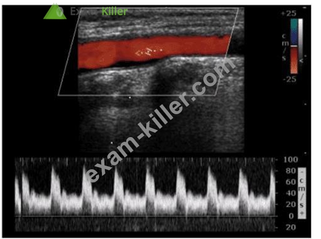
- A. Decreasing velocity scale
- B. Increasing sweep speed
- C. Decreasing Doppler gain
- D. Increasing the gate size
Answer: C
Explanation:
Noise in the Doppler waveform can often be attributed to excessive gain settings. Decreasing the Doppler gain reduces the amplification of both the signal and the noise, thus providing a clearer and more accurate Doppler waveform. Excessive gain can cause speckling and clutter, which obscure the true Doppler signals.
By reducing the gain, the noise level is minimized, resulting in a cleaner Doppler signal representation.
References:
ARDMS Sonography Principles & Instrumentation Guidelines
Hagen-Ansert SL. Textbook of Diagnostic Ultrasonography. 8th ed. St. Louis, MO: Mosby; 2017.
NEW QUESTION # 37
......
Download Exam SPI Practice Test Questions with 100% Verified Answers: https://www.exam-killer.com/SPI-valid-questions.html
Share Latest SPITest Practice Test Questions, Exam Dumps: https://drive.google.com/open?id=1qzKGXs2qR6uizfOHblMb6860pahZXdjn